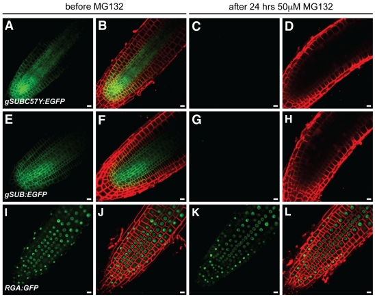
Figure 11. Effects of MG132 treatments on the expression of different SUB::gSUB:EGFP reporters.
Live confocal microscopy images from roots were generated using 4-day old Arabidopsis seedlings (Ler) carrying different SUB:EGFP reporters. The same root is shown before (A–B, E–F, I–J) and after (C–D, G–H, K–L) 24-hrs treatment with 50 µM MG132. The FM4-64 stain was used to mark the outline of all cells in a tissue. The signals from the EGFP and FM4-64 channels are shown in green and red, respectively. (A–D) SUB::gSUBC57Y:EGFP. (E–H) SUB::gSUB:EGFP. (I–L) A RGA::RGA:GFP reporter that served as control [93]. Note that signal persisted after MG132 treatment (K–L). Scale bars: 10 µm.

