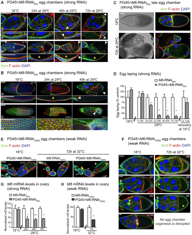
Figure 6. M6 knockdown in the follicular epithelium impairs oogenesis.
PG45>M6-RNAi flies were raised at 18°C and then adults were either kept at 18°C (control) or transferred to 29°C or 32°C for 24, 48 or 72 hours to induce the M6-RNAi in the FE. Ovaries were dissected and stained with phalloidin (red), DAPI (blue) and Armadillo (green; A–C, E–F). Confocal images of PG45>M6-RNAiNIG (PG45; tub-GAL80ts; UAS-M6-RNAiNIG) egg chambers corresponding to early and mid-oogenesis (A) and late oogenesis (B–C) are shown. Gaps and loss of FE organization and integrity are indicated by white arrowheads. Note defects in the FC shape (yellow asterisks, *). (B) After a 72 hour induction of M6 knockdown, egg chambers reaching late oogenesis could not be assigned due to gross morphological defects. Two views of a st11 egg chamber incubated at 29°C for 24 hours and 48 hours are presented in (a, 0 µm) and (b, 11.26 µm). (a′–b′) Magnified views of the indicated regions. Supplementary information is presented in Fig. S8. (C) Light transmission and fluorescent images of late egg chambers incubated at 18°C (upper panel) and 29°C for 72 hours (lower panel). Note the aberrant morphology induced by M6 downregulation. Scale bar is 50 µm. (D) Egg laying was determined as in Figure 3A. PG45>M6-RNAiNIG (grey bars) and control females, without the PG45 driver (white bars), were incubated at 18°C and transferred to 29°C. After 76 hours at 29°C, flies were allowed to recover at 18°C for 144–168 hours and the phenotype was restored. Mean ± s.e.m., n = 2; Mann Whitney test between genotypes at each condition, p<0.05 (*). (E–F) Confocal images of egg chambers from PG45>M6-RNAiVDRC (PG45; UAS-M6-RNAiVDRC; tub-GAL80ts) females incubated at 18°C or 32°C for 72 h are shown. (E) Arrested ovarioles were detected in PG45>M6-RNAiVDRC incubated at 32°C for 72 h, which were never evidenced in controls. The white asterisk indicates an arrested st9 egg chamber. Scale bar is 50 µm. (F) No late st9 was observed in PG45>M6-RNAiVDRC induced at 32°C for 72 h. Yellow asterisks point to defects in follicle cell shape, which are not as columnar as control, whereas green arrowheads indicate the position of polar and border cells (st9). Note that at 32°C the cluster of border and polar cells did not migrate posteriorly in late st9 egg chambers. Scale bar is 20 µm. (G–H) M6 mRNA levels were measured in ovaries by RT-qPCR. (G) Ovaries from PG45>M6-RNAiNIG (grey bars) and control females, without the PG45 driver (white bars), were dissected from adult females incubated at 18°C or at 29°C for 24, 48 and 72 hours. Mean ± s.e.m., n = 3; unpaired t-tests with Welch correction between genotypes, p>0.05 at 18°C; p<0.05 at 29°C for 24 hours and 72 hours (*); p<0.01 at 29°C for 48 hours (**). (H) Ovaries from PG45>M6-RNAiVDRC (dark grey bars) and control females, without the PG45 driver (white bars), were dissected from adult females incubated at 18°C or at 32°C for 72 hours. Mean ± s.e.m., n = 3; unpaired t-test with Welch correction between genotypes, p>0.05 at 18°C, p<0.05 at 32°C (*).

