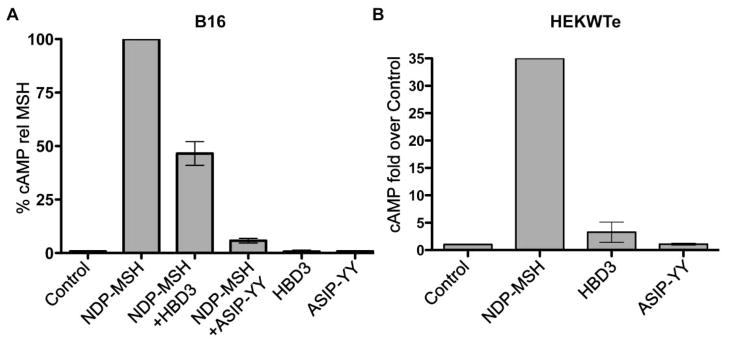
Figure 2.
cAMP accumulation in B16 or HEK293 cells stably transfected with melanocortin MC1 receptor (HEKWTe). Cells were incubated in serum-free media for at least 2 hours. Cells were pre-incubated with 0.1mM IBMX for 15min, then stimulated with the indicated ligands for 10min. cAMP levels were quantified with the cAMP EIA system (Amersham Biosciences). The bars indicate the range from two independent experiments
A) 0.5nM or 1nM NDP-MSH alone or in combination with 100nM agouti signaling protein peptide (ASIP-YY) (McNulty et al., 2005) or HBD3, or 100nM ASIP-YY or HBD3 alone. Data was normalised to NDP-MSH, which was set at 100%. Results are from two independent experiments.
B) 1nM NDP-MSH, 100nM HBD3 or 100nM ASIP-YY. Data was expressed as fold over the control. Results are from 2 independent experiments, except for NDP-MSH, which is from only one experiment.

