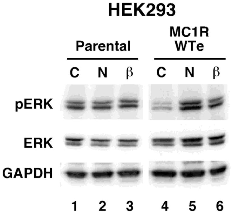
Figure 3.

ERK phosphorylation in response to α-melanocyte stimulating hormone and human β-Defensin 3. Lanes are as indicated C=Control, N=10nM NDP-MSH, β=10nM HBD3. HEK293 untransfected cells (parental) or HEK293 stably expressing melanocortin MC 1 receptor (WTe) were pre-incubated in serum-free media for at least 2 hours, then stimulated with the indicated ligands for 5min. Western immuno-blotting with the indicated antibodies was performed using total cell extracts. pERK (Cell Signalling) is specific to the phosphorylated Thr202/Tyr204 sites of ERK1/2, ERK (Cell Signalling) recognises all forms of the ERK protein. Anti-GAPDH (R&D Systems) was used as a loading control. This blot is representative of three independent experiments.
