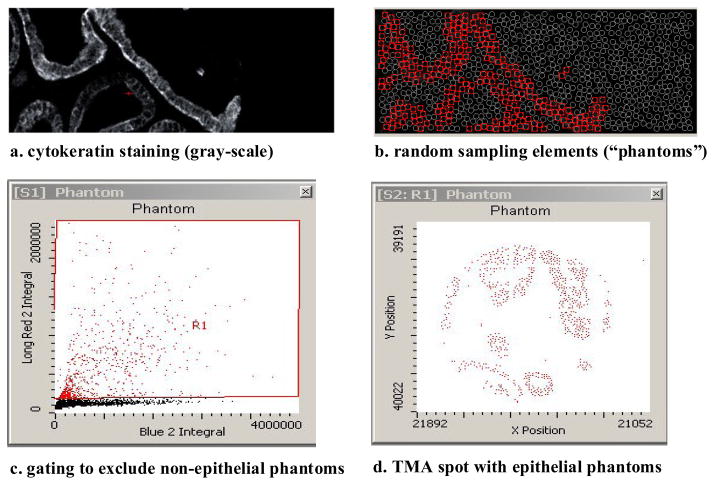
Figure 1.
Detection of epithelial areas using stereological “phantoms”, a dense array of circles randomly placed on the image. Each phantom becomes a distinct object that can be sorted according to its content of cytoplasm (cytokeratin), nucleus (DAPI) and p27. Phantoms containing little or no epithelium are discarded by gating in a scatterplot.

