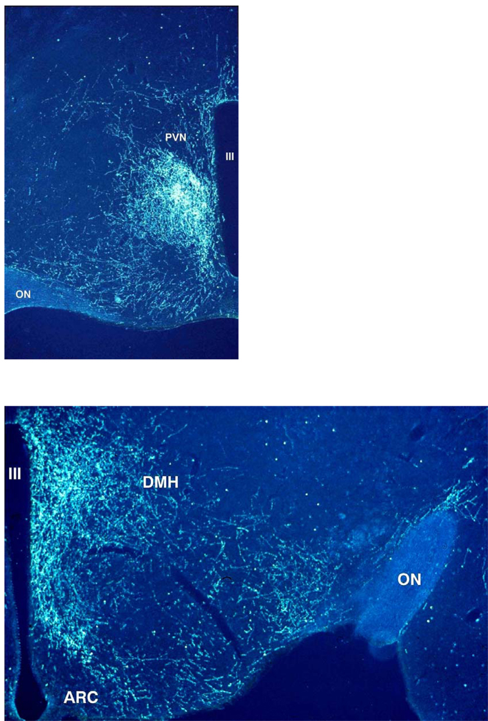
Fig.1. Darkfield illustration of SCN projections into the hypothalamic paraventricular (PVN), dorsomedial (DMH) and ventromedial (VMH) hypothalamus.
After the iontophoretic injection of Phaseolus vulgaris leucoagglutinin (PHA-L) into the SCN a large concentration of labelled fibers can be observed in the area just ventral to the PVN (upper panel), also known as the sub-paraventricular zone or the subPVN. Less dense but also clearly innervated in the upper panel are the periventricular part and the dorsomedial part of the PVN, but note the almost complete lack of labelled fibers in the parvicellular and magnocellular parts of the PVN. Caudal to the PVN (lower panel) a dense terminal field of SCN fibers can be observed in the DMH. In the lower panel also a clear concentration of PHA-L labelled fibers is present in the area between the arcuate nucleus (ARC) and the VMH, as well as the area lateral to the VMH. The third ventricle (III) is on the right side (upper panel) or left side (lower panel).

