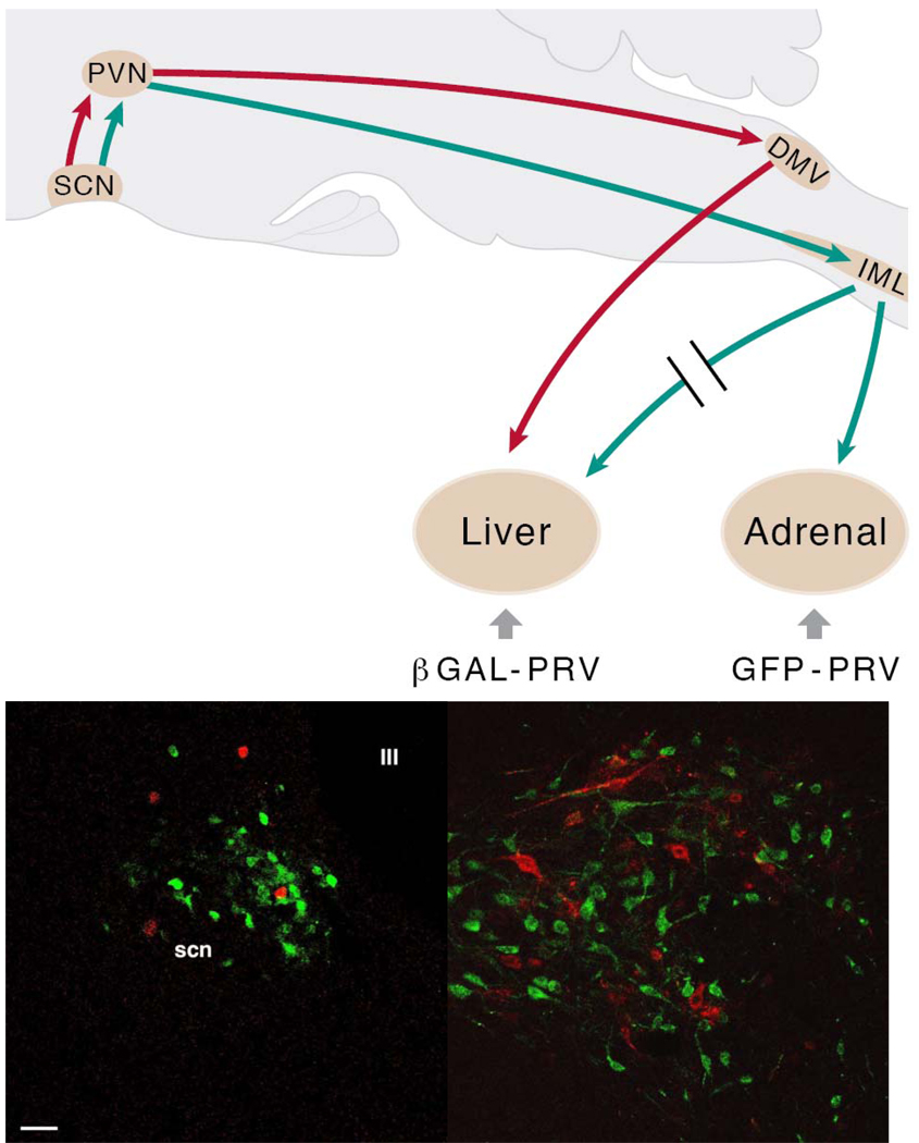
Fig.5. The suprachiasmatic nucleus (SCN) balances sympathetic and parasympathetic output to peripheral organs through separate pre-autonomic neurons.
In the upper panel the experimental setup used to examine the possible separation of sympathetic and parasympathetic pre-autonomic neurons in the hypothalamus is indicated in a schematic drawing of a sagittal section of the rat brain. B-galactoside PRV (βGAL-PRV) was injected into the sympathetic denervated liver, forcing the virus to infect the brain via the vagus nerve (red lines); simultaneously the pre-sympathetic neurons were labelled by an injection of green fluorescent protein (GFP)-PRV into the adrenal (green lines). After the labelling of the first-order neurons in the brainstem and spinal cord, this approach resulted in separate pre-parasympathetic and sympathetic neurons in the PVN (second order), followed by a similar separation of the third-order neurons in the SCN. In the lower panels transverse sections of the hypothalamus in the region of the PVN (left) and SCN (right) show a perfect separation of pre-parasympathetic βGAL-PRV (red) and pre-sympathetic GFP-PRV (green) labelled neurons, as there are no yellow (i.e., double-labelled) neurons. Scale bar = 25 µm. With permission adapted from (Buijs et al., 2003).

