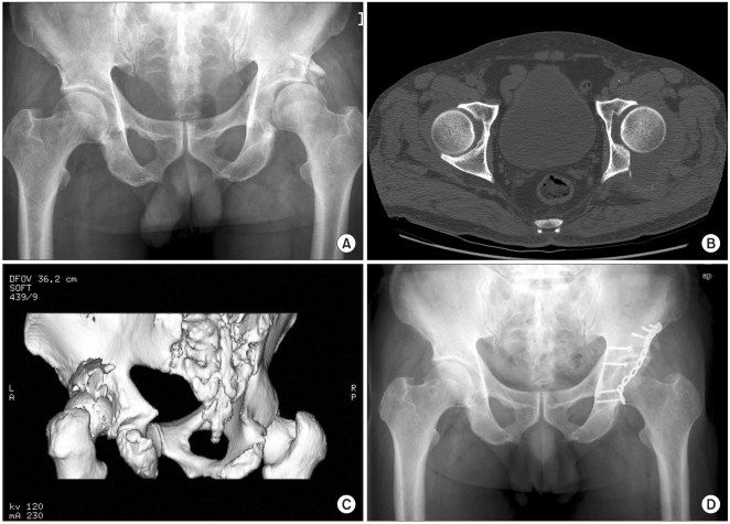
Fig. 1.
(A) Preoperative radiograph and (B) 2-dimensional and (C) 3-dimensional CT scans of a 65-year-old male with posterior wall fracture. After reduction of the osteochondral fragments with an autologous bone graft into the bone void, we performed internal fixation of the fracture with 2.4 mm lag screws and a reconstruction plate. (D) radiograph taken 3 years after surgery, the patient was free of pain with an adequate range of motion.

