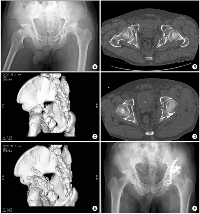
Fig. 2.
(A) Preoperative radiograph and (B) 2-dimensional and (C) 3-dimensional CT scans of a 46-year-old male with posterior wall fracture associated with a transverse acetabular fracture. We performed reduction of the osteochondral fragments with an autologous bone graft and internal fixation of the fractures with 2.0 mm mini-screws, a spring plate and two reconstruction plates. (D) Two-dimensional and (E) 3-dimensional CT scans after surgery show congruous reduction of the posterior wall. (F) Radiograph taken 2 years after surgery, the patient showed excellent functional results.

