Figure 1.
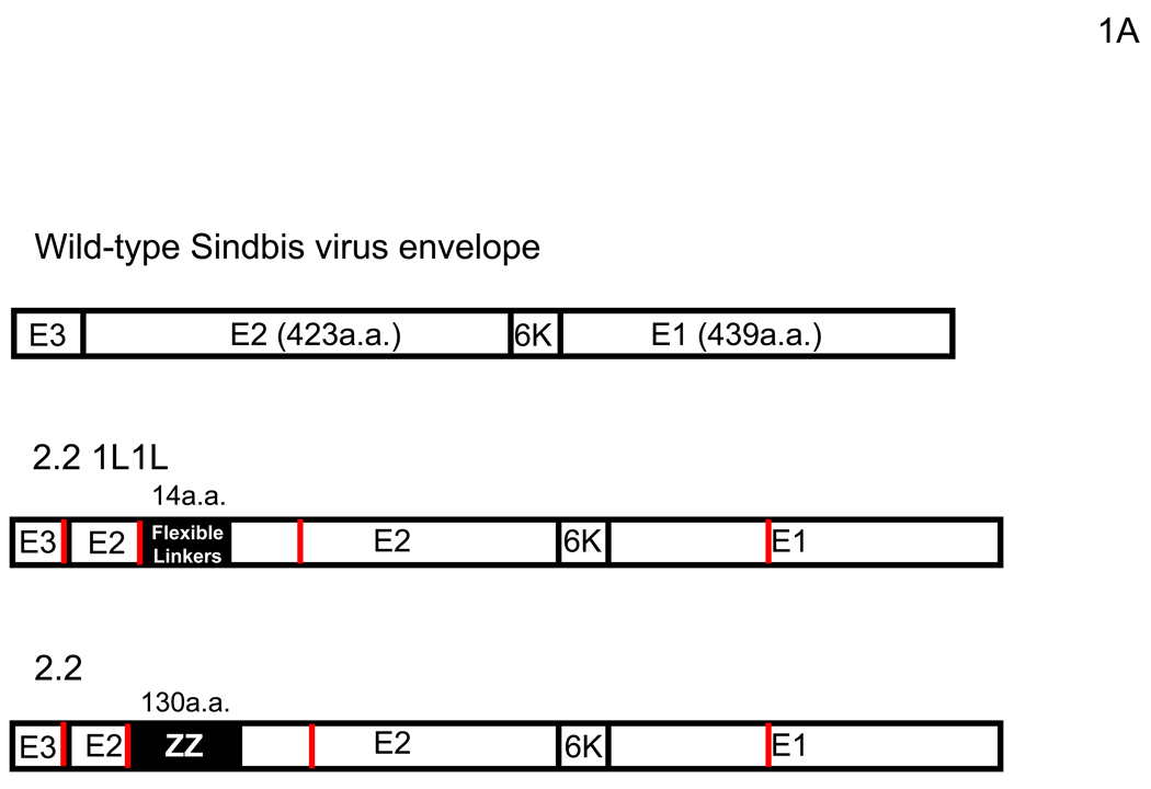
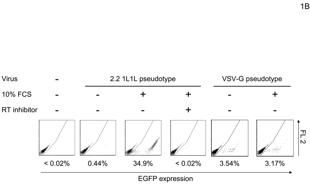
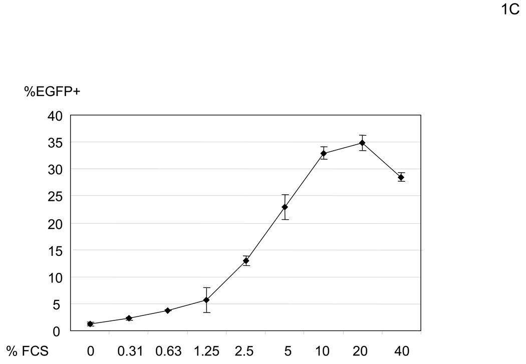
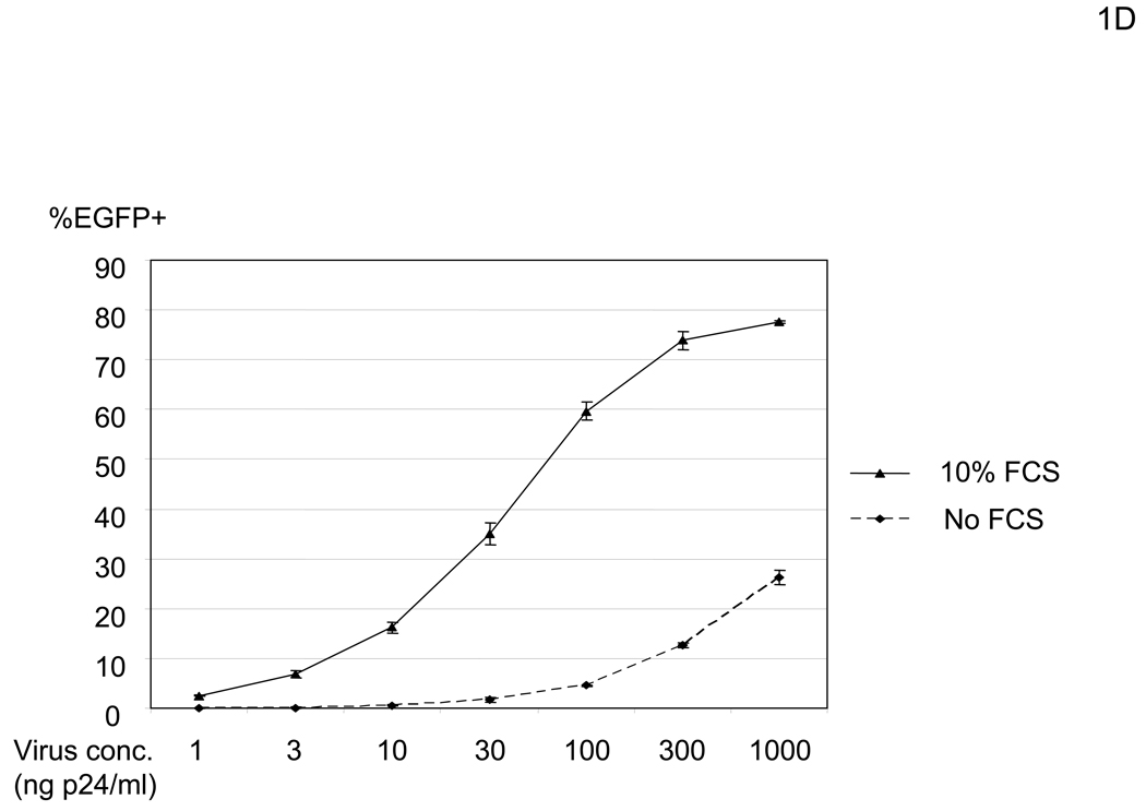
FCS enhances transduction with lentiviral vectors pseudotyped with various types of envelope proteins. (A) Schematic representation of chimeric Sindbis virus envelope proteins. The Sindbis virus envelope protein is first synthesized as a polypeptide and subsequently cleaved by cellular proteases to generate the E3, E2, 6K, and E1 proteins. E1 and E2 are incorporated into the viral envelope. Both 2.2 and 2.2 1L1L were derived from the wild-type Sindbis virus envelope protein with mutations (shown as red lines) to reduce their binding to original receptors. In addition, 2.2 1L1L has two flexible linkers and 2.2 contains the ZZ domain at the E2 amino acid 70. (B) For all transduction and infection experiments, 1×10⁴ HMVEC/well were seeded into 48-well plates 2 days before infection or transduction. Total infection/transduction volume was 150 µl. HMVEC were incubated with PBS (+) or 10% FCS in PBS (+), followed by transduction with 2.2 1L1L (100 ng p24/ml). Nevirapine (5 µM) was used 30 minutes before, during, and after transduction. VSV-G-pseudotype (10 ng p24/ml) was also used instead of the 2.2 1L1L pseudotype. (C) HMVEC were incubated with different concentrations of FCS in PBS (+) and transduced with the 2.2 1L1L pseudotype (100 ng p24/ml). (D) The enhancing effects of 10% FCS on HMVEC transduction were investigated, using various concentrations of 2.2 1L1L pseudotype. In this manuscript, all error bars and the values after ± represent standard deviations of triplicated experiments.


