Figure 5.
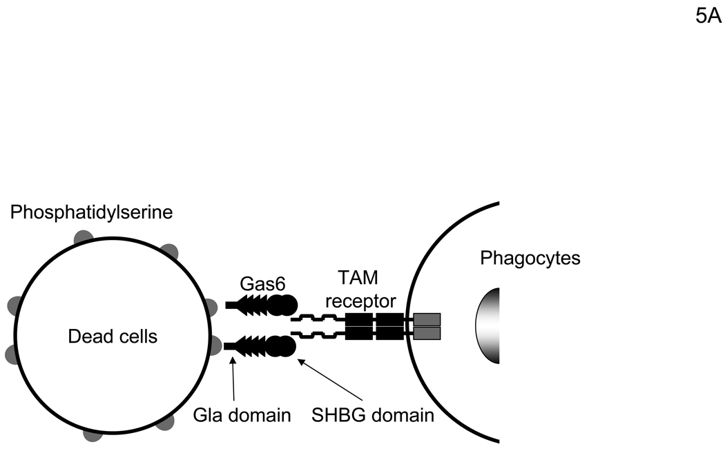
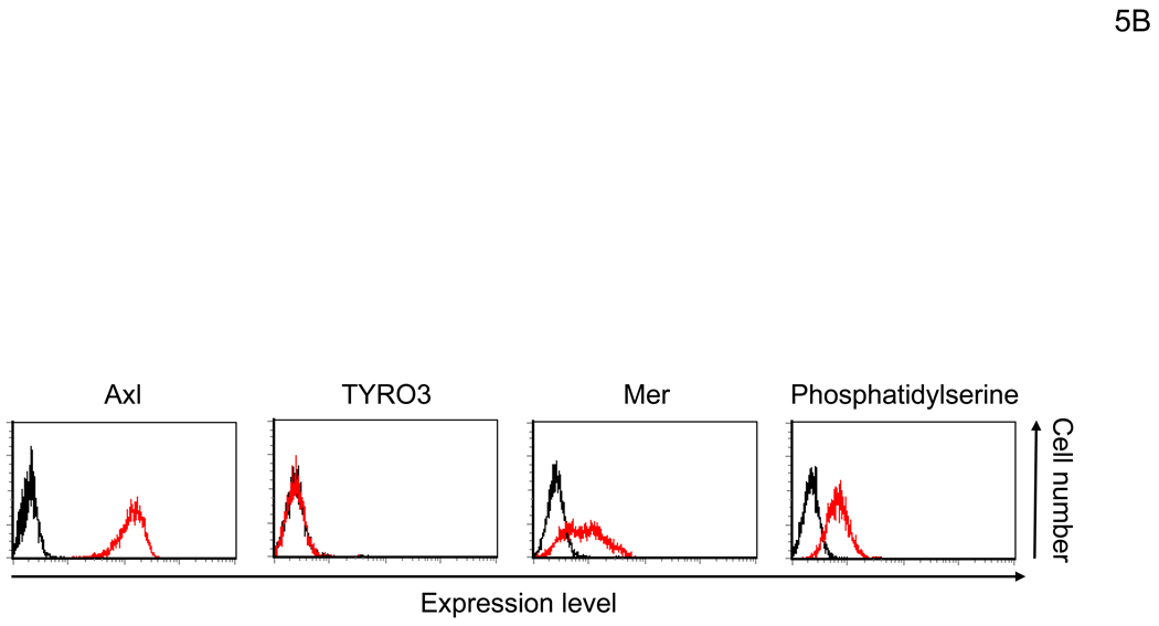
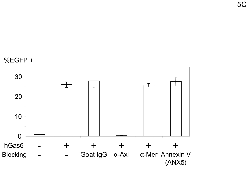
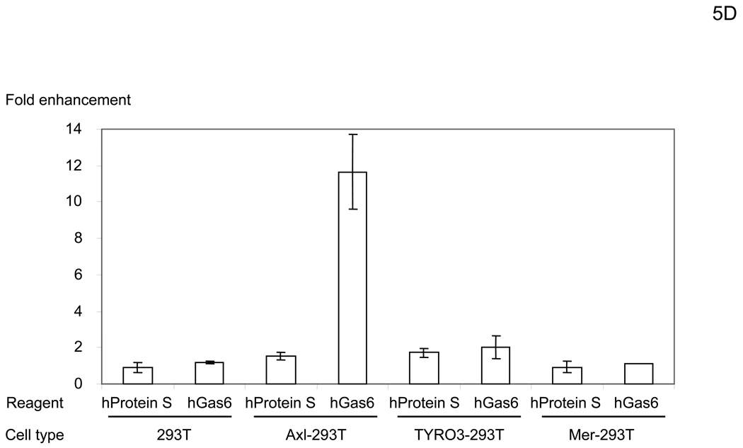
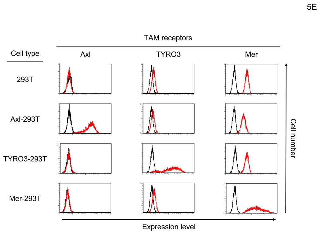
hGas6 binds to Axl on target cells. (A) Schematic representation of the mechanism whereby hGas6 mediates phagocytosis of dead cells. (B) Expression of Axl, TYRO3, Mer, and PtdSer on HMVEC. (C) HMVEC were incubated for 1 hour with PBS (+) or PBS (+) containing one blocking reagent (control IgG, anti-human Axl antibody, anti-human Mer antibody, or ANX5) (25 µg/ml) then incubated with hGas6 (100 ng/ml) in the presence of each blocking reagent for 4 hours. After removing hGas6 and blocking reagents, the cells were transduced with the 2.2 1L1L pseudotype (100 ng p24/ml). (D) Enhancement of viral transduction of 293T, Axl-293T, TYRO3-293T, and Mer-293T cells mediated by hGas6 or hProtein S. 1×10⁵ cells were incubated with PBS (+), PBS (+) containing hProtein S, or hGas6 (100 ng /ml), followed by transduction with the 2.2 1L1L pseudotype (100 ng p24/ml). The fold enhancement was calculated by the following formula: % of EGFP expressing cells preincubated with hProtein S or hGas6 divided by % of EGFP expressing cells preincubated with PBS (+). (E) Expression of Axl, TYRO3, and Mer on 293T, Axl-293T, TYRO3-293T, and Mer-293T cells. Black and red lines are staining with isotype control antibodies and antibodies against each type of TAM receptor, respectively.

