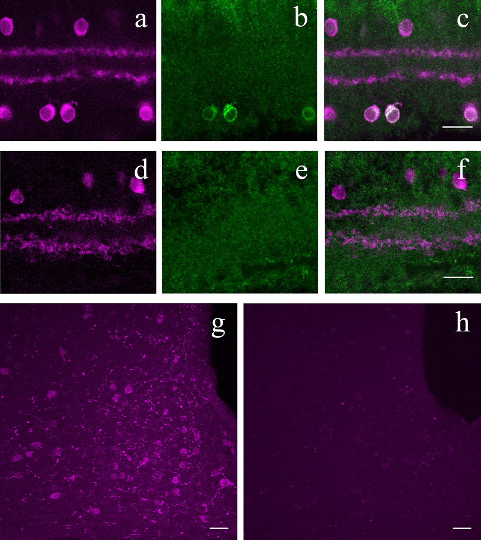
Figure 10.
β-endorphin antibody labeling is specific to POMC neurons in both retina and hypothalamus. A: POMC-DsRed (magenta) retina showing DsRed+ soma distribution in both the INL and GCL with two DsRed bands in the IPL. B: Same region as in A, showing β-endorphin+ somas within GCL. C: A merged image of A and B, showing colocalization of POMC-DsRed+ cell bodies with β-endorphin+ somas. D: Vertical cryostat section through POMC-KO retina immunolabeled for ChAT (magenta), showing ChAT+ cell bodies, with two bands in the IPL. E: Image illustrating the same area as D, stained with anti-bodies against β-endorphin (green), showing no specific labeling of somas or projections. F: A merged image of D and E, showing only ChAT+ cell bodies and projections. G: β-endorphin immunolabeling (magenta) in the arcuate nucleus of hypothalamus in wild type mouse. H: β-endorphin immunolabeling (magenta) in the arcuate nucleus of the hypothalamus in POMC KO (Pomc−/−Tg) mouse. Scale bars: 20µm.

