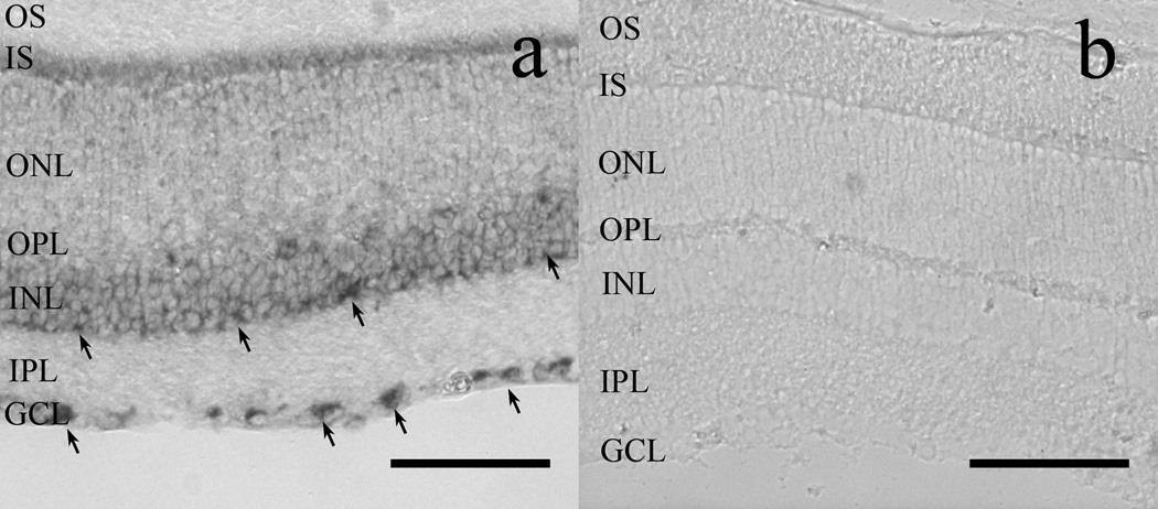
Figure 8.
In situ hybridization reveals POMC mRNA in the GCL and INL of wild-type mouse retinas (A). Note the dark reaction product obtained with the antisense probe, indicative of POMC mRNA expression in somas located in the GCL and in INL (arrows). (B): The sense probe failed to label any structure in the retina. OS: photoreceptor outer segment layer; IS: photoreceptor inner segment layer; ONL: outer nuclear layer; OPL: outer plexiform layer; INL: inner nuclear layer; IPL: inner plexiform layer; GCL: ganglion cell layer. Scale bars: 80µm.

