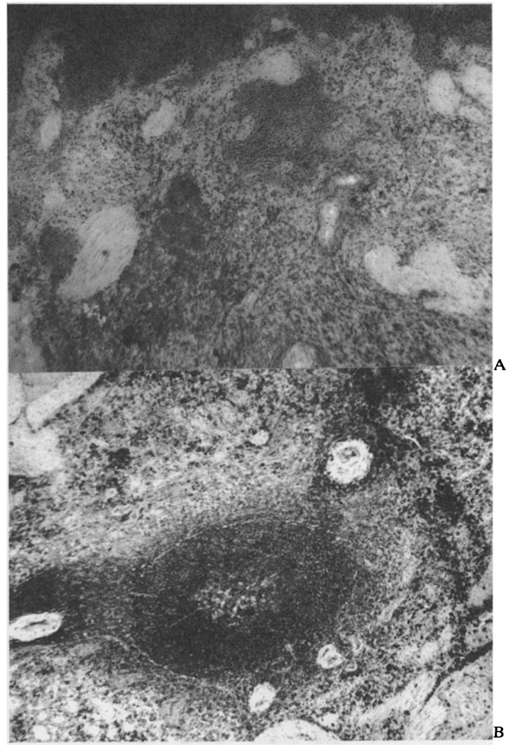
FIGURE 4.
a. Photomicrograph of autografted spleen at nine days. Note the scattered hemorrhage. Moderate numbers of pyroninophilic cells are present in the red pulp. The white pulp is decreased and germinal centers are absent (methyl-green-pyronin stain, × 80).
b. Photomicrograph of splenic autograft at eight months. Note the well-developed white pulp. Numerous pyroninophilic cells are present throughout (methyl-green-pyronine stain, × 80).

