
Figure 3. MEK Is Essential for DNA Damage-Induced Checkpoint Response.
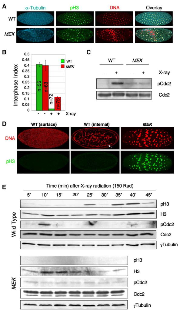
(A) Embryos (0–1 hr AED) were treated with low-dose X-ray radiation (150 rad), allowed to develop for 35 min at 25°C, and then fixed and stained with propidium iodide (red) and antibodies for α-Tubulin (cyan) and phospho-Histone H3 (pH3; green).
(B) Interphase index of embryos 35 min following the treatment in (A). The interphase index is defined as the percentage of pH3-negative embryos of the total. “n” denotes the number of embryos counted. Error bars represent standard deviation.
(C) Immediately following X-ray irradiation (within 10 min) as in (A), embryo lysates were subjected to SDS-PAGE and blotted sequentially with indicated antibodies.
(D) 60 min following irradiation as in (A), embryos were fixed and stained with propidium iodide (red) and anti-pH3 (green). The arrow points to a second nuclear layer formed by falling nuclei (middle). Note the unusually large and fragmented DNA (pH3-positive) in the MEK embryo.
(E) Freshly laid eggs (0–1 hr AED) of indicated genotypes were treated with 150 rad X-ray, and then ten embryos were homogenized at each indicated time point. Embryo lysates were subjected to SDS-PAGE and blotted sequentially with indicated antibodies. The membrane was stripped of antibodies between blotting.