Figure 1.
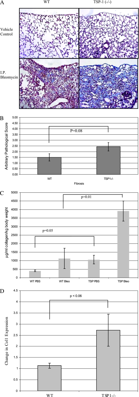
Analysis of bleomycin-induced fibrosis in thrombospondin-1 (TSP-1)−/− mice. (A) TSP-1−/− mice suffer more fibrosis than do wild-type (WT) animals after intraperitoneal (I.P.) bleomycin challenge. Representative trichrome stains of these lungs are shown. Data are representative of six mice per group. (B) TSP-1–deficient mice tended to manifest more fibrosis after bleomycin challenge. A certified veterinary pathologist blindly scored each slide for fibrosis. The average pathology scores ± SEMs for six mice per group are shown. No fibrosis was evident in vehicle-treated control mice (data not shown). (C) TSP-1−/− lungs contain more collagen according to Sircol assay after bleomycin challenge. Lungs were homogenized with acetic acid, and Sircol dye was added to each sample to bind collagen. Alkali reagent was added to release the bound dye into solution, and absorbance at a 540-nm wavelength was obtained. Results were normalized to the dead weight of each mouse. The average data ± SEMs are shown for a minimum of four animals per treatment group. P = 0.05, comparing bleomycin-treated WT mice (WT Bleo) versus TSP-1–deficient mice. (D) Bleomycin-challenged TSP-1−/− mice exhibited more collagen I mRNA than did WT animals in whole-lung preparations after bleomycin treatment. RNA was extracted and subjected to real-time RT-PCR analysis, using SYBR Green. Shown is the average fold increase of Collagen I (Col1) expression over Glyceraldehyde 3-phosphate dehydrogenase (GAPDH) ± SEMs in untreated versus bleomycin-treated mice.

