Figure 1.
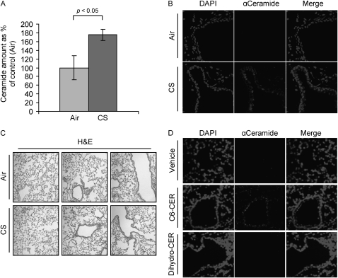
Cigarette smoke (CS) induces ceramide generation in the lung. Three 129/Sv mice for each time/treatment point were exposed, or not, to CS for 1 week (A and B), whereas two 129/Sv mice for each treatment/point were intratracheally instilled with ceramide analogs or BSA (vehicle) (D). The mice were killed and assayed for ceramide levels in the lung by diacylglycerol kinase assay (A) and immunohistochemistry (IHC) (B and D). (A) The amount of ceramide in the graphic represents the average of three independent experiments (three mice per treatment), and is reported as percentage of the untreated mice (filtered air) after normalization per protein unit of the tissue; the P value in (A) was obtained by Student's t test per number of mice. (B) Total nuclei (left panels) were stained by 4′,6-diamidino-2-phenylindole (DAPI ceramide (central panels) was localized by incubating lung slides with a specific anti- (α) ceramide antibody (Ab) and stained by Alexa Fluor 555 dye–conjugated Ab; the panels on the right show the merges of total nuclei and ceramide staining. (C) Hematoxylin and eosin (H&E) stain of the 129/Sv mice exposed, or not, to CS for 1 week. (D) mice were instilled with synthetic lipids, either C6-ceramide (C6-CER) or dihydro-C6-ceramide (Dihydro-CER), killed 24 hours after instillation, and assayed for ceramide levels in the lung by IHC: ceramide (central panels) was localized as in (A) and stained by Alexa Fluor 555 dye; total nuclei were stained by DAPI (left panels); merge images of ceramide and nuclei staining are shown (right panels). Images were acquired with an LSM 5 Pascal Zeiss laser scanning or an Olympus FluoView FV1000 confocal microscope.

