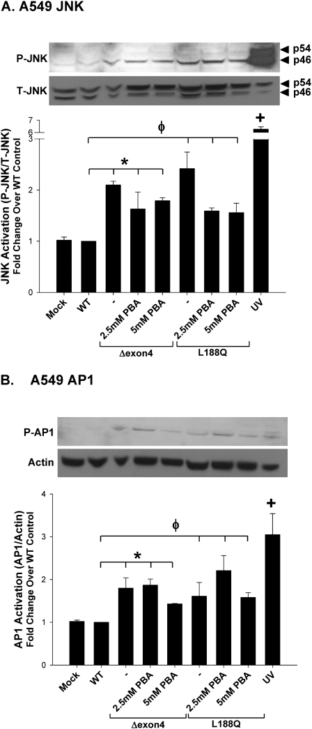
Figure 8.
SP-C mutant induction of JNK and AP-1 is not altered by 4-PBA treatment. A549 cells were transiently transfected in the absence (Mock) or presence of the indicated EGFP–SP-C constructs, and harvested 48 hours after transfection. Eight hours after transfection, increasing concentrations of 4-PBA were added to SP-CΔexon4–transfected cells and harvested 48 hours after transfection. UV treatment (40 mJ/cm2) was used for a positive control sample. Cell lysates were analyzed for phospho-SAPK/JNK and total SAPK/JNK expression (A) and phospho–c-Jun (AP1) expression (B). An anti–β-actin antibody was used as a loading control. Band intensities were quantified by densitometry, and the ratio of either phospho-JNK to total JNK, or c-Jun to β-actin, was determined. Data are expressed as fold change over WT control, and represent the mean ± SD of three separate experiments. Representative immunoblots appear above each graph. *P < 0.05 for SP-CΔexon4 samples versus WT control. φP < 0.05 for SP-CL188Q samples versus WT control. +P < 0.01 for UV positive control versus WT control.

