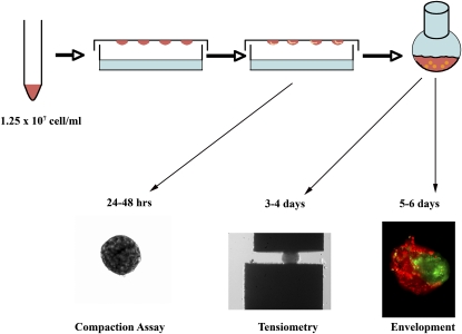
Figure 1.
Fetal pulmonary cells in three-dimensional (3D) suspension self-assemble to form pulmonary bodies (PBs). Fetal lungs isolated at Embryologic Day 14.5 were enzymatically dissociated and resuspended in 3D hanging drops (HDs). Pulmonary cells (1.25 × 10⁷ cell/ml) self-assembled or compacted over 48 hours to form pulmonary sheets (compaction assay). Pulmonary sheets placed in a shaker flask for 24–48 hours formed spherical PBs. These were subjected to tissue surface tensiometry (TST) to measure aggregate cohesivity (tensiometry), or to envelopment assays in which pairs of differentially stained PBs were apposed in 3D HDs and examined by fluorescence microscopy after 24–48 hours (envelopment).

