Figure 2.
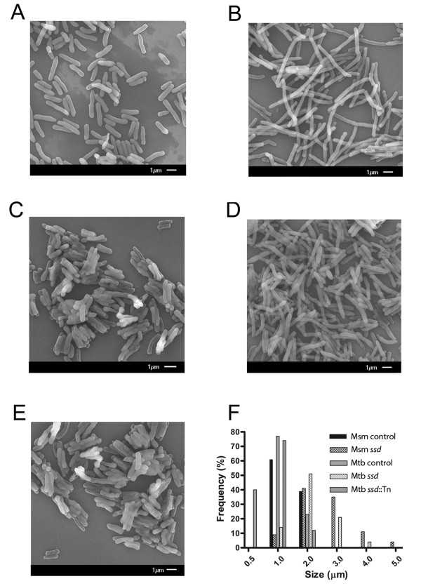
Ultrastructure Analysis (SEM) and Length distributions. Bacterial morphology. (A) M. smegmatis control strain, (B) M. smegmatis ssd merodiploid (C) M. tuberculosis control, (D) M. tuberculosis ssd merodiploid and (E) ssd::Tn mutant M. tuberculosis strain were visualized by scanning electron microscopy. Images are representative of different fields of bacteria from exponentially growing cultures at 37°C. (F) Lengths of the bacterial cells were calculated from the coordinates of both ends of the cell as measured from representative fields as visualized by scanning electron microscopy. Multiple fields were examined and values calculated in 0.5-1 mm increments from multiple fields of over 100 cells.

