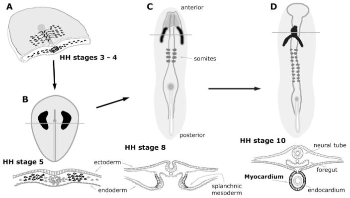
Fig. 1.
Schematic diagram depicting the initial morphological events in the development of the avian heart. Precardiac and definitive myocardial tissue are illustrated in black, while the lines overlaying the cardiac areas denote the plane of the transverse sections shown immediately below the embryos. A: Three-dimensional view of a cross-sectioned chick embryo, which is in transition between HH stages 3 and 4. Precardiac cells undergo gastrulation through the primitive streak and move laterally to reside in lateral mesoderm within the anterior half of the embryo. B: During HH stage 5, the myocardial progenitors coalesce into morphologically distinct heart-forming fields, which are distributed bilaterally to the primitive streak. C: At HH stage 8, the two heart-forming areas have sorted to splanchnic mesoderm and begun to merge at the embryonic midline. By this stage, these cells have become definitive cardiac myocytes, as they will exhibit a number of muscle proteins in a prestriated pattern. D: By HH stage 10 a fully contractile heart has developed. This primitive heart tube consists of an outer sheet of myocardium, surrounding an inner endocardial layer.

