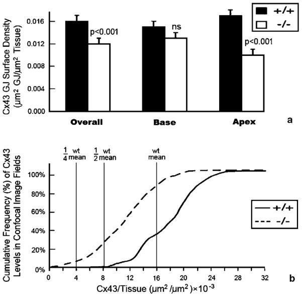
Fig. 2.
Quantitative analyses of spatial heterogeneities in ventricular Cx43 in HF-1b knockout myocardium. (a) Cx43 content (μm2) per unit area of ventricular myocardium (μm2), shows an overall decrease in the HF-1b knockout ventricle compared to wildtype littermates. This reduction in Cx43 content is mainly localized to the apical territory of the ventricle (p <0.001). (b) Cumulative frequency distributions of Cx43 content measurements taken from the 90 optical sections sampled within wildtype and knockout apical myocardium. The leftward shift of the knockout plot demonstrates a population Cx43 immunolabeled optical sections with extremely low Cx43 content levels not found in the wildtype.

