Fig. 3.

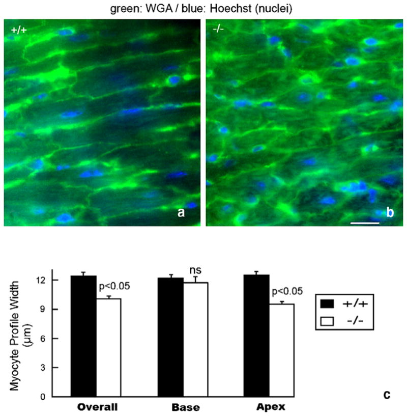
Diminished myocyte diameter and morphologic irregularity in the HF-1b knockout ventricular myocardium (a, b) WGA–FITC (green) and Hoechst (blue) staining delineate myocyte sarcolemma and nuclei in the apical myocardium of wildtype (a) and knockout (b) ventricles. (c) Plots of mean myocyte diameter overall in the ventricle and within the basal and apical territories. Significant differences (p <0.05) between knockout and wildtype measurements were found. Scale bar=25 μm.
