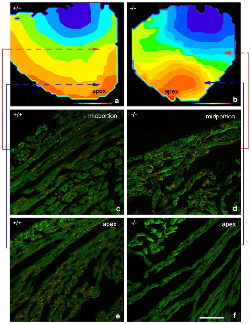
Fig. 5.

Direct co-localization of an apically located sector of disrupted and reduced Cx43 and a region of slow conduction in the HF-1b knockout ventricle. Isochronal maps of knockout and wildtype ventricles are shown in (a) and (b), respectively. (c–f) Cx43 immunolabeling (red) in ventricular regions indicated by shaded squares on the isochronal maps. Colored lines indicate shaded areas corresponding to areas of Cx43 immunolabeling. Note the reduction in Cx43 content in the apical region of the knockout ventricle (f), corresponding precisely with an area of slowed conduction in (b).
