
Figure 2.
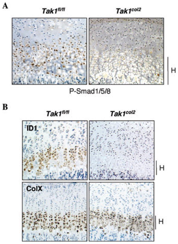
Reduced activation of BMP-responsive SMADs in TAK1col2 mice in vivo. (A) Immunohistochemically stained sections showing phosphorylation of BMP-responsive Smad proteins. Coronal sections of the proximal tibia of P0 Tak1fl/fl and Tak1col2 pups were stained with anti-phospho-Smad1/5/8 antibody. Hypertrophic chondrocytes in the terminal growth plate are indicated with an H. (B) In situ hybridization for ID1 and Collagen Xα (ColX). Coronal sections of the proximal tibia of 3-week-old Tak1fl/fl and Tak1col2 mice were probed for the expression of the indicated mR-NAs. The hypertrophic region of the growth plate is shown.