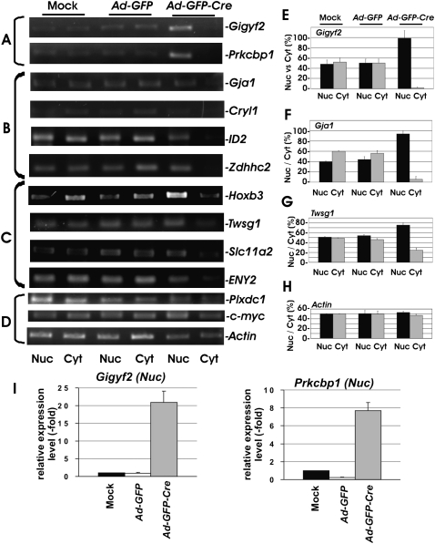
FIGURE 2.
Down-regulation of mRNA export by the depletion of THOC5 in fibroblasts. MEF THOC5 flox/flox cells were infected with adenovirus carrying the GFP gene (Ad-GFP) or adenovirus carrying the GFP and Cre-recombinase genes (Ad-GFP-Cre). Four days after infection, nuclear (Nuc) and cytoplasmic (Cyt) RNAs were isolated and applied for RT-PCR using primers as described in Materials and Methods. cDNA from all samples were standardized by an equal level of actin mRNA in both fractions. We have performed three to five independent experiments, and we show one example of representative data (A–D). (A) mRNAs were accumulated in the nucleus and were not exported. (B) mRNAs were not accumulated in the nucleus and also were not exported. (C) mRNAs were not accumulated in the nucleus, and mRNA export was reduced. (D) There was no significant alteration by the depletion of THOC5. (E–H) Signal intensity from Gigyf2- (E), Gja1- (F), Twsg1- (G), and actin-specific (H) RT-PCR products was quantified using TINA 2.0 software. The percent signal intensity from the nuclear (Nuc) or the cytoplasmic (Cyt) fraction of total intensity (Nuc + Cyt) in Mock MEF THOC5 flox/flox (Mock) or MEF THOC5 flox/flox infected with Ad-GFP or Ad-GFP-Cre. Average values ±SD from three independent experiments are shown. (I) Aliquots of cDNA samples from nuclear fractions of A were applied for quantitative RT-PCR analysis of Gigyf2 and Prkcbp1 mRNA. Relative expression levels compared to HPRT1 were normalized to gene expression in mock-infected MEF THOC5 flox/flox. Average values from four independent PCR reactions ±SEM are shown.

