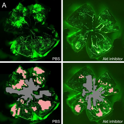
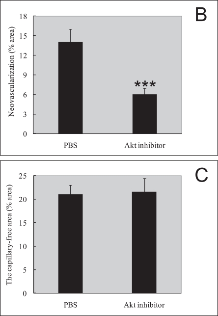
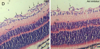
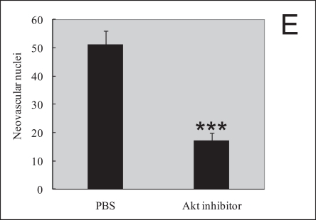
Fig. 3.
Akt inhibitor treatment suppressed retinal neovascularization but not vessel loss in the murine oxygen-induced retinopathy model. (A) Representative retinal whole-mounts showing area of capillary-free and neovascularization after 5 days of relative oxygen deficiency exposure and intravitreal injections (P12) of Akt inhibitor and PBS. Areas of capillary (gray) and neovascularization (pink) were quantified. Original magnification ×5. (B) The areas of neovascularization and the total areas were quantified. Data are shown as mean±SEM. (PBS, n=10; recombinant Akt, n=15; ***P≤0.001). (C) The capillary-free areas and the total areas were quantified. (D) Histologic sections of P17 retina from PBS-treated (left) and Akt inhibitor-treated (right) eyes of mouse exposed to hyperoxia. Extensive preretinal neovascular tufts were apparent (arrow). (E) The number of neovascular nuclei anterior to the ILM in 6-µm retinal sections was quantified. (PBS, n=10; recombinant Akt, n=15; ***P≤0.001).


