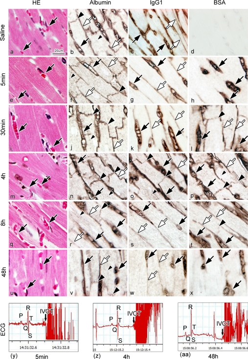
Fig. 6.
Light micrographs of HE-stained heart sections with IVCT after saline injection (a) or at various times after the BSA injection (e, i, m, q, u). Serial sections are immunostained for albumin (b, f, j, n, r, v), IgG1 (c, g, k, o, s, w), and BSA (d, h, l, p, t, x). After the saline injection (a–d), the BSA immunostaining is not detected (d). At 5 min after the BSA injection (e–h), BSA is immunolocalized only in blood vessels (h; black arrows). At 30 min (i–l), BSA is immunolocalized in blood vessels (l; black arrows), interstitium (l; white arrows), and intercalated discs (l; large black arrowhead), in the same pattern as the albumin immunolocalization (j). At 48 hr (u–x), BSA is immunolocalized in blood vessels (x; black arrows), but not in intercalated discs and interstitium. At 4 hr (m–p), IgG1 is immunolocalized in intercalated discs (o; large black arrowheads), blood vessels (o; black arrows), and interstitium (o; white arrow). At 8 hr (q–t), IgG1 is only immunolocalized in blood vessels (s; black arrows) and interstitium (s; white arrows). (y)–(aa) Arrows in ECG show the exact times of freezing hearts with IVCT at 5 min (y), 4 hr (z), and 48 hr (aa) after the BSA injection, showing early diastolic phases. Bar=20 µm.

