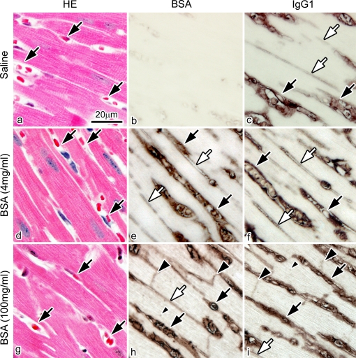
Fig. 9.
Light micrographs of serial sections of mouse heart tissues prepared by IVCT at 4 hr after the saline injection or at two concentrations of 4 mg/ml or 100 mg/ml BSA injection. The serial sections are stained with hematoxylin-eosin (HE; a, d, g) or immunostained for BSA (b, e, h) and IgG1 (c, f, i). (b) There is no immunopositive staining for BSA at 4 hr after the saline injection. (e), (f) The BSA and IgG1 are immunolocalized only in blood vessels (black arrows) and interstitium (white arrows) at 4 hr at the concentration of 4 mg/ml BSA. (h), (i) However, they are immunolocalized in the intercalated discs (large black arrowheads) and t-tubules (small black arrowhead) in addition to blood vessels (black arrows) and interstitium (white arrow) at the concentration of 100 mg/ml. Bar=20 µm.

