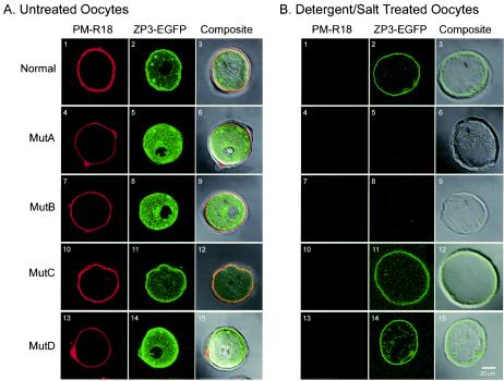
FIG. 4.
Confocal microscopy of mutant ZP3-EGFP expressed in growing oocytes. Plasmid vectors expressing normal, MutA, MutB, MutC, or MutD ZP3-EGFP were injected into the nucleus of growing oocytes and cultured for 40 h. Oocytes were incubated with a lipid membrane stain (PM-R18) before (A1, 4, 7, 10, and 13) or after (B1, 4, 7, 10, and 13) freeze-thawing in the presence of 0.5 M NaCl and 1% NP-40. PM-18 (A1, 4, 7, 10, and 13 and B1, 4, 7, 10, and 13) and ZP3-EGFP (A2, 5, 8, 11, and 14 and B2, 5, 8, 11, and 14) were viewed by individually and as a composite (A3, 6, 9, 12, and 16 and B3, 6, 9, 12, and 15) after superimposition on a light microscopic image. Scale bar, 20 μm.

