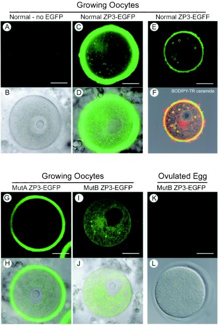
FIG. 5.
Confocal microscopic imaging of ZP3-EGFP mutants expressed in transgenic mice. Mouse lines with transgenes expressing either normal, MutA, or MutB ZP3-EGFP were established and compared to normal, nontransgenic mice. Almost fully grown oocytes or eggs were isolated and imaged by confocal microscopy to detect EGFP alone (A, C, G, I, and K) or superimposed on images obtained by differential interference contrast optics (B, D, H, J, and L). No background signal was detected in the cytoplasm or zona pellucida of normal mice (A and B), and the strongest signal was observed in fully grown oocytes from normal ZP3-EGFP transgenic mice (C and D). In smaller growing oocytes, normal ZP3-EGFP also was observed in large circular structures (E) that costained with BODIPY-TR ceramide (F). (G and H) A diminished, although significant signal, was observed in MutA ZP3-EGFP mice. (I and J) No EGFP was observed in the zona pellucida of MutB ZP3-EGFP mice, but reporter protein was present in the cytoplasm and incorporated in the circular structures. (K and L) EGFP was not observed in the cytoplasm or in the zona pellucida of ovulated eggs isolated from MutB ZP3-EGFP transgenic mice. Scale bar, 20 μm.

