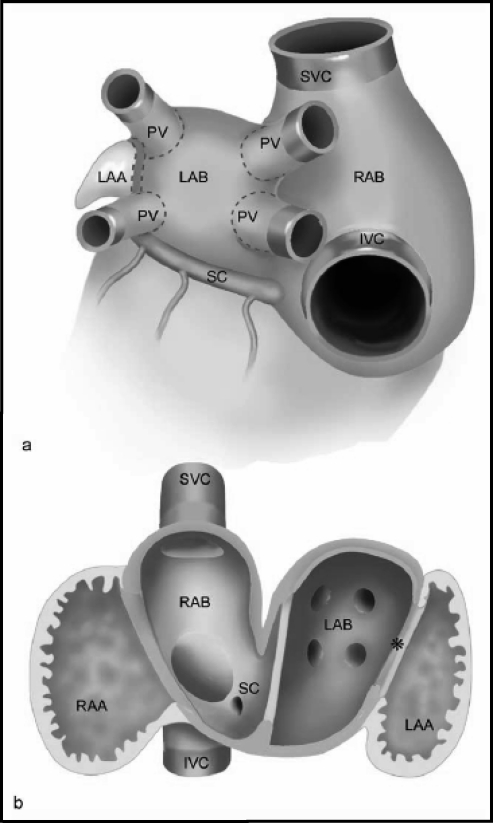
Figure 2.
(a) Schematic depicting the outer sides of the atrial chambers with pulmonary veins (PV), superior vena cava (SVC), and inferior venal cava (IVC). The right and left atrial bodies (RAB and LAB) are covered by smooth-walled inner myocardium that stretches into the extracardiac PVs and partially into the systemic veins. (b) Schematic depicting the tissue seen from inside the left and right atria and how the smooth-walled myocardial tissue extends into the systemic veins. Left-sided sinus venosus tissue (*) and SC = coronary sinus.
(Reprinted with permission from Douglas YL, Jongbloed MR, Gittenberger-de Groot AD, et al. Am J Cardiol. 2006;97:662–670.18)

