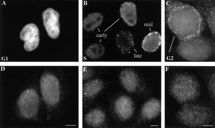
Figure 3.
HeLa cells at several different stages of interphase contain RNA foci from replication-dependent histone genes. (A) A pair of mid-G1 daughter cell nuclei, 4 h after mitotic shake-off, is counterstained with DAPI. (B) In an asynchronous population of HeLa cell nuclei pulse-labeled with BrdU, anti-BrdU antibody staining reveals patterns indicative of cells in early, middle, and late S phase. (C) Immunostaining with anti-cyclin B antibody reveals one G2 cell with cytoplasmic cyclin B particularly concentrated around the nuclear periphery (top left) and one cell with only background signal, not in G2 (lower right). (D–F) H4 RNA foci are detected by FISH in the same cells shown in A–C. In these examples, RNA foci are clearly visible in both G1 cells (D), one of the early S phase cells (E), and the G2 nucleus (F). Typically, early S phase cells have the brightest and most numerous RNA foci (E), though RNA has been detected in all stages of S phase. Bars, 5 μm.

