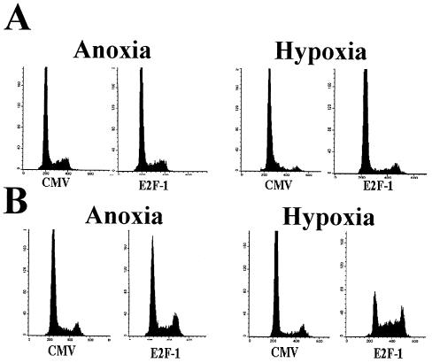
FIG. 8.
Effects of E2F expression on proliferation of anoxic and hypoxic cells. (A) REF52 cells were rendered anoxic (left) or hypoxic (right) for 24 h and then infected with either CMV or E2F-1 adenovirus. After an additional 24 h, the cells were collected and analyzed for DNA content. (B) Cells were serum starved for 48 h and then rendered hypoxic (right) or anoxic (left). The cells were then stimulated with serum and infected with either CMV or E2F-1; 24 h later, the cells were collected and analyzed for DNA content.

