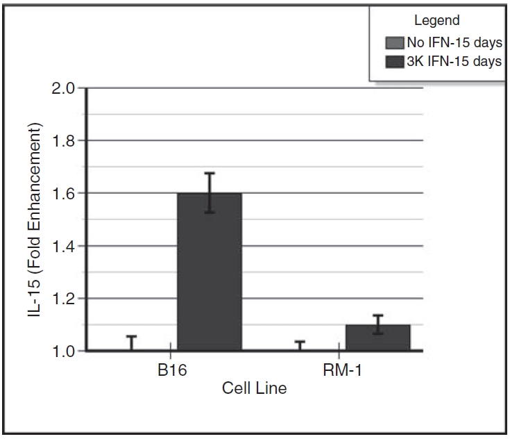
FIG. 3. .

Induction of membrane-associated interleukin-15 (IL-15) in interferon-α (IFN-α)–treated B16α cells and RM-1α cells. Monolayers of B16α and RM-1α cells were grown in medium containing 3,000 units/mL of IFN-α for 15 days. Control monolayers of parental cells were given medium alone without IFN treatment. At 2 days after providing fresh medium with or without IFN-α, the cell monolayers were assayed for membrane-bound IL-15 using a whole-cell ELISA. Standard curves of IL-15 were run concomitantly with the samples. The data are expressed as IL-15 fold enhancement (mean ± SE) versus days of IFN treatment. ANOVA analysis for membrane-associated IL-15: day 16 B16 parental versus day 16 B16α cells: P < 0.0001; day 16 RM-1 parental cells versus day 16 RM-1α cells: P = NS.
