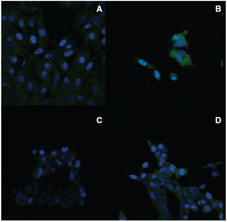
FIG. 4. .

Subcellular distribution of interleukin-15 (IL-15) protein in B16α and RM-1α cells. Parental cells and interferon (IFN)-treated α-cells were cultured in the absence or presence of 3,000 IU/mL of IFN-α for 2 weeks, respectively. The cells were fixed, permeabilized, and incubated with or without (data not shown) primary antibody to IL-15 and subsequently incubated with secondary antibody to IgG conjugated with fluorescein isothiocyanate (green fluorescence). Nuclei were stained with DAPI (blue fluorescence). Fluorescence was visualized with a Zeiss LSM510 UV META confocal microscope. The illustrations represent overlays of the blue and green fluorescence images. All images have the same magnification with the scale. (A) B16 parental cells incubated with primary antibody to IL-15. (B) B16α cells incubated with primary antibody to IL-15. (C) RM-1 parental cells incubated with primary antibody to IL-15. (D) RM-1α vaccine cells incubated with primary antibody to IL-15.
