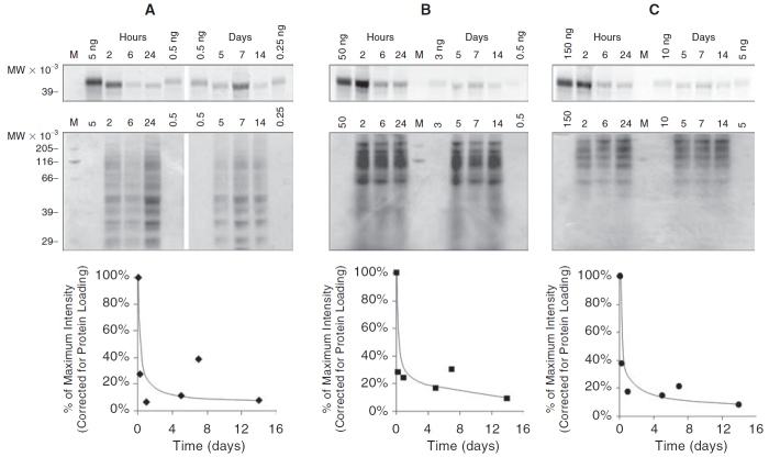
FIG. 6. .
LDS-PAGE of eye tissue extracts from in vivo experiments. In brief, 20 μL of ReGel at 2 mg Alexa 647 ovalbumin/mL ReGel (40 μg Alexa ovalbumin total) were injected into rat eyes and after indicated periods of time, tissues were dissected from eyes, protein was extracted, resolved by LDS-PAGE, and compared to Alexa 647 ovalbumin gel standards. For each tissue type, retina (A), choroid/RPE (B), and sclera (C), the gels were prepared and scanned for fluorescence (Typhoon) to indicate the migration of ovalbumin that was extracted from each tissue (top narrow row of gel images). The gels were later stained with Coomassie Blue to determine total protein loading on the gels for each lane (larger middle row of gel images). The intensity of the Typhoon fluorescent signal was determined, corrected for protein loading, normalized to the maximum value and plotted over time from 2 h to 14 days (bottom row of graphs).

