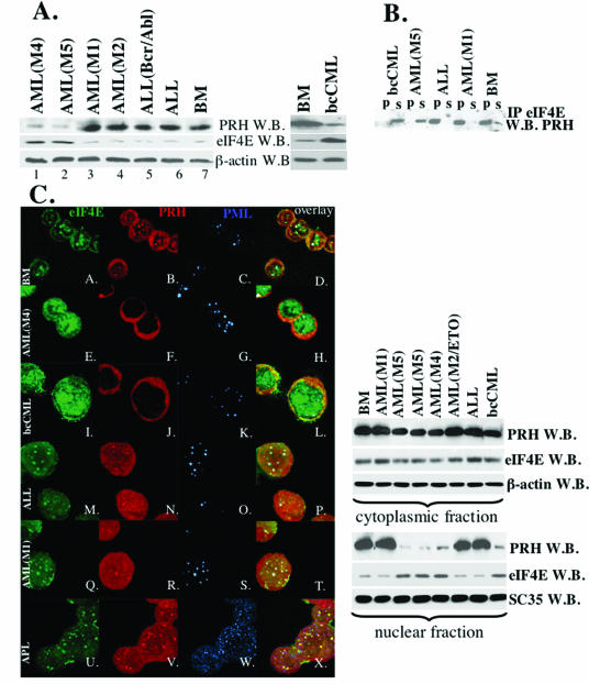
FIG. 1.
Levels, subcellular distribution, and interaction of eIF4E and PRH are altered in M4 AML, M5 AML, and bcCML specimens. (A) Western blot analysis of whole-cell extracts in cells derived from specimens as indicated. β-Actin is shown as a control for protein loading. (B) Whole-cell lysates were immunoprecipitated with anti-eIF4E Ab (IP eIF4E), and the resulting Western blot was probed for PRH. IP, immunoprecipitated fraction; s, supernatant after immunoprecipitation; W.B., Western blot. (C) In the left panel are shown confocal micrographs of cells stained with FITC-conjugated anti-eIF4E Ab (shown in green); anti-PRH Ab, followed by Texas red-conjugated anti-rabbit IgG Ab (shown in red); and anti-PML Ab (5E10), followed by Cy5-conjugated anti-mouse IgG Ab (shown in blue). The PML-eIF4E overlay is shown in light blue, the PML-PRH overlay is shown in pink, the PRH-eIF4E overlay is shown in yellow, and the triple eIF4E-PML-PRH overlay is shown in white. The objective was 100x with a further magnification of 2 (A-H) or 3 (I-X) fold. In the right panel, cells were fractionated into cytoplasmic and nuclear compartments and analyzed as indicated. β-Actin and SC 35 were used as a loading control for the cytoplasmic and nuclear fractions, respectively. BM, cells derived from the healthy individuals. Other specimens are as indicated. AML-ETO indicates that translocation was found in that M2 specimen (Table 1).

