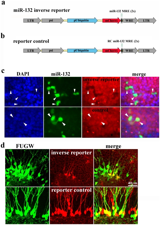
Figure 1. An Inverse Reporter for Detecting miR-132 Expression In Vivo.
a, We generated an inverse miR-132 reporter lentivirus by placing two perfectly complementary miR-132 target sequences (miR-132 MRE) downstream of mCherry driven by an internal ubiquitin promoter (pUbiquitin). This inverse reporter design results in suppression of mCherry expression in the presence of miR-132. b, The reporter control virus was generated by placing the reverse complement of the miR-132 target (RC miR-132 MRE) downstream of mCherry. c, Hek293 cells were infected with either the inverse reporter or reporter control lentivirus. Single cells were cultivated to generate clonal cell lines expressing the miR-132 inverse reporter (top row) or the inverse reporter control (bottom row). Infection of a proportion of the cells with a miR-132-expressing retrovirus (green) suppressed mCherry in the inverse reporter cells (black silhouettes in red panel, top row), but not in inverse reporter control cells (red panel, bottom row). d, We co-injected equal titers of the GFP-expressing FUGW virus and either the mCherry-expressing miR-132 inverse reporter virus or the reporter control virus into 8 week old mice. FUGW lentivirus showed widespread infection of granule cells at 7 days post-injection (DPI, left panels). In contrast, the mCherry expressing miR-132 inverse reporter virus primarily labeled cells along the subgranular zone of the dentate gyrus (top row, middle panel). The merge is shown in the right panel. Infection of the reporter control virus showed the same widespread infection as the GFP-expressing FUGW virus (lower row, left and middle panel).

