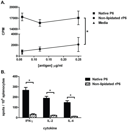
Figure 3. Lipid motif on P6 augments T cell proliferation and cytokine production.
WT mice were immunized s.c. with 40 µg of native P6 emulsified in CFA and IFA one week later. (A) Proliferation of CD3+ cells isolated from draining lymph nodes was measured following 4 day co-culture with syngeneic irradiated BMDCs pulsed with 0.25, 0.12, and 0.06 µg/ml native P6 (▪) or non-lipidated rP6 (•). Media alone control (▴) was performed for background proliferation of T cells. Thymidine was added to the wells for the last 16 hrs of incubation. (B) Splenocytes from the same animals were co-cultured overnight with 0.06 µg/ml antigen pulsed irradiated BMDCs in ELISPOT plates coated with anti-cytokine mAb. Plates were developed and spots enumerated microscopically. *p<0.01 1way ANOVA with Bonferroni post-test comparison of native P6 to non-lipidated rP6.

