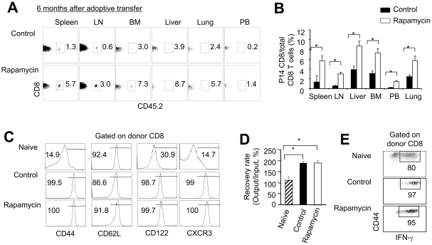
Figure 6. Rapamycin T cells become long-lived memory T cells in vivo.
P14 CD44loCD8+ TN were stimulated in vitro with B6-derived DCs pulsed with gp33 plus IL-2, in the absence or presence of rapamycin (100 nM). Six days later, T cells were recovered, washed and adoptively transferred into sublethally irradiated Thy1.1 B6 mice. Donor T cells were collected from these Thy1.1 B6 recipient 6 months after transfer. Graphs show the percentage (A) and number (B) of P14 CD8+ derived CD8+ T cells recovered from different organs. (C) Histograms show the expression of indicated surface markers on P14 naïve T cells and donor CD8 T cells recovered from mice receiving rapamycin T cells and mice receiving control T cells. (D and E) P14 CD8+Thy1.2+ cells (1×10⁴) were highly purified using cell sorter and were stimulated in vitro with B6 DCs pulsed with gp33 (10−13 M) for another 8 days. P14 CD8+ naïve T cells stimulated with B6 DCs pulsed with gp33 (10−13 M) for 6 days were analyzed as controls. Cells were recovered from the cultures and counted. Graphs show the number of cells (D). Dot plots show the fraction of IFN-γ producing cells (E). Data are shown as means ± SD and representative of two independently performed experiments with 3 mice in each group. *p<0.05, significant difference.

