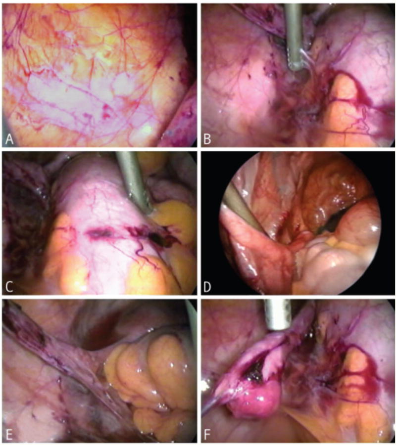
Figure 1. Laparoscopic visualization of endometrial lesions (A – F).

(A)Isolated red lesions located on broad ligament. (B)Multiple red lesions adjacent to sigmoid colon. Laparoscopic instrument visualized in the top center portion of image. Lesions visualized directly to left of instrument. (C)Hemorrhagic lesion located on sigmoid colon in the center of the image. (D)Hemorrhagic lesion on broad ligament posterior to round ligament. Lesion is located immediately to right of laparoscopic instrument. (E)Adhesions formed at base of oviduct adjacent to the uterus. Hemorrhagic endometrial lesions located at left of image with filmy adhesions. (F)Filmy adhesions and hemorrhagic lesion located at distal portion of oviduct.
