
Figure 3.
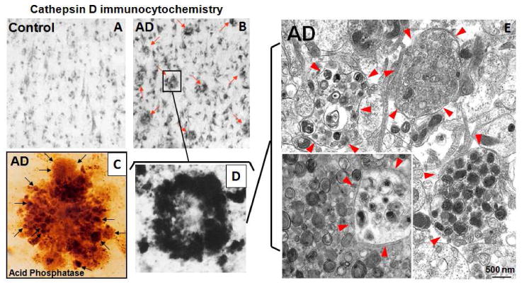
The “burden” of AVs containing incompletely digested proteins stored in dystrophic neurites of the AD brain is great. At the light microscopic levels, CatD immunostaining in the AD brain (B) but not in the control brain (A) reveals a high density of senile plaques (B, arrows) each containing dozens of dystrophic neurites visible after histochemical staining for acid phosphatase (C, arrows) or by CatD immunocytochemistry (D). (E) Ultrastructural analysis of dystrophic neurites (circled by arrowheads) demonstrates that dystrophic neurites predominantly contain hundreds of AVs of distinct subtypes but the majority have double membranes with electron dense lumens, most consistent with autolysosomes and amphisomes in which proteolysis of substrates, including inner membrane, has stalled. Given the number of dystrophic neurites in the AD brain, the amount of “stored” wasted proteins is enormous, rivaling that in certain primary lysosomal storage disorders.