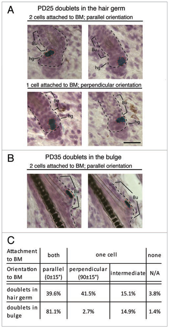
Figure 2.
Placement of two bulge daughter cells with respect to the niche. Skin sections (20 µm) from K14CreER × Rosa26R mice (injected with tamoxifen at PD17 and stained for X-Gal and hematoxylin at days indicated) show doublet orientation with respect to the basement membrane (BM) and in two distinct compartmets: the differentiating zone (hair germ) and the stem cell niche (bulge). (A) Bulge cells exported in the germ divided both parallel (top) and perpendicular (bottom) with respect to the BM. (B) Bulge cells that continue to reside in the niche divided parallel to the BM. (C) Quantification of data in (A and B). BM, black dotted lines; Bu, bulge; hg, hair germ. Scale bar, 50 µm. N = 127 hair follicles.

