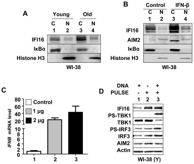
FIGURE 6.
IFI16 protein is primarily detected in the cytoplasm of old or IFN-β treated HDFs and nucleofection of young HDFs with dsDNA induces the IFN-β expression. A, Cultures of young (PD ~30) proliferating or old (PD ~50) WI-38 HDFs were harvested and cells were subjected to the nuclear and cytoplasmic fractionation. The fractions containing equal amounts of proteins were analyzed by immunoblotting using antibodies specific to the indicated proteins. B, Cultures of young proliferating WI-38 HDFs were either left untreated or treated with IFN-β for 24 h. After the treatment, cells were subjected to the nuclear and cytoplasmic fractionation. The fractions containing approximately equal amounts of proteins were analyzed by immunoblotting. C, Young proliferating (PD ~30) WI-38 HDFs were nucleofected without DNA (control) or with the indicated amounts of plasmid DNA (GFP plasmid) as described in methods. After 18 h of nucleofection, total RNA was extracted and analyzed by quantitative real-time PCR for steady-state levels of IFNB mRNA. The ratio between the IFNB mRNA levels to actin mRNA was calculated in units (one unit being the ratio of the IFNB mRNA to actin mRNA). The relative steady-state levels of IFNB mRNA in control HDFs are indicated as 1. D, Young proliferating WI-38 HDFs were either left without nucleofection or nucleofected without DNA or nucleofected with 2 μg of plasmid DNA. After 18 h of nucleofection, total cell lysates were analyzed by immunoblotting for the indicated proteins.

