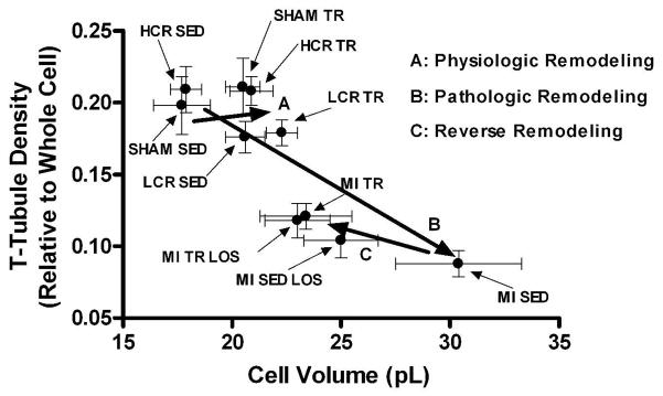
Figure 5.
The relationship between cardiomyocyte volume and transverse (T)-tubule density in cardiomyocytes from sham-operated (SHAM) or post-myocardial infarction (MI) heart failure rats and rats with either a high or low capacity for running (HCR and LCR, respectively) that remained either sedentary (SED) or underwent exercise training (TR). LCR and post-MI heart failure rats developed respectively mild concentric and severe eccentric/concentric pathologic remodeling. Moreover, SED and TR post-MI heart failure rats that also received the angiotensin II type 1 receptor antagonist losartan were also included. Note that the cardiomyocyte volume-T-tubule density relationship is right-shifted after TR-induced physiologic hypertrophy in normal and mild cardiac pathology, but left-shifted after TR-induced reverse remodeling in post-MI heart failure rats with severe cardiac pathology, indicating that T-tubule density at least partly is dissociated from cell size. Data are mean±SEM with 8 animals/group.

