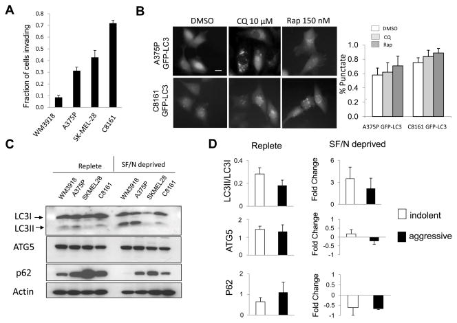
Figure 2. Invasiveness and tumor cell autophagy in aggressive and indolent melanoma cell lines.
(A) Invasion rate by the Boyden Chamber assay (B) Autophagy modulation with the autophagy inhibitor chloroquine (CQ) or the autophagy inducer rapamycin (Rap) in cell lines expressing the autophagy marker GFP-LC3. Diffuse fluorescence: no autophagy; punctate fluorescence: accumulation of autophagic vesicles. (C) Immunoblotting against autophagy markers in aggressive (SKMEL28, C8161) and indolent (WM3918, A375P) cell lines grown in complete medium:Replete; and in serum free (24 hours) and nutrient free medium (2 hours):SF/N deprived. (D) Gel densitometry of autophagy markers in replete and SF/N deprived media. Fold change of markers in SF/N deprived conditions were compared to Replete measurements.

