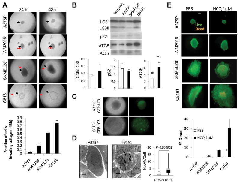
Figure 4. Growth, invasion, and tumor cell autophagy of aggressive and indolent melanoma spheroids grown in a three dimensional collagen matrix.
(A) Brightfield microscopy of spheroids; black arrow: central spheroid boundary; red arrow: boundary of invasion (B) immunoblots of lysates derived from spheroids 48 hours after implantation in collagen; mean +/− SD gel densities for indolent (WM3918, A375) and aggressive (SKMEL28, C8161). (C) Brightfield and fluorescent microscopy of A375P GFP-LC3 and C8161GFP-LC3 spheroids (48 hours) (D) Electron micrographs of A375P and C8161 spheroids (48hours); arrows: autophagic vesicles. (D) Live (green)/Dead (orange) assay of melanoma spheroids in 3D culture 48 hours after the indicated treatments, HCQ: hydroxychloroquine; Scale bar: 200 μm (A,C,E); 2 μm (D).

