
Figure 1. Impact of S6k1 genetic deletion on lymph nodes proliferation and pheochromocytoma formation triggered by heterozygous loss of Pten.
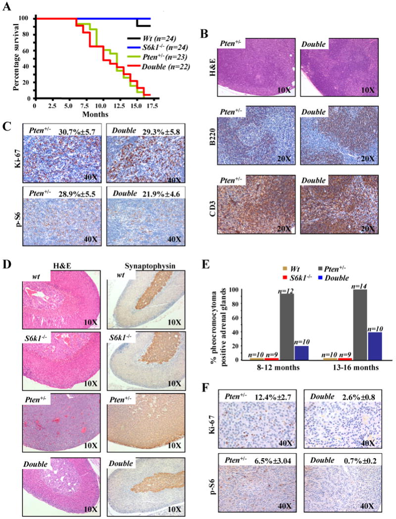
A, Kaplan-Meier plot for overall survival shows no difference between S61k-/-;Pten+/- (double) and Pten+/- mice. B, sections of lymph nodes from 12 month old S6k1-/-;Pten+/- and Pten+/- mice stained with: haematoxylin and eosin (H&E) (upper panel); anti-B220, a specific marker of B-lymphocytes (middle panel); anti-CD3, a specific marker of T-lymphocytes (lower panel). C, sections of lymph nodes from 12 month old S6k1-/-;Pten+/- and Pten+/- mice stained with: Ki-67 (upper panel); anti-phospho-S6 (lower panel). Quantifications of Ki-67 and phospho-S6 positive cells are indicated. D, sections of adrenal glands from 10 month old wt, S6k1-/-, Pten+/- and S6k1-/-;Pten+/- mice stained with: H&E (left panel) and anti-synaptophysin, a specific marker of the medulla of the adrenal gland (right panel). E, cumulative incidence of pheochromocytoma over time in wt, S6k1-/-, S6k1-/-;Pten+/- and Pten+/- mice. F, sections of adrenal gland from 10 month old S6k1-/-;Pten+/- and Pten+/- mice stained with: Ki-67 (upper panel); anti-phospho-S6 (lower panel). Quantifications of Ki-67 and phospho-S6 positive cells are indicated.