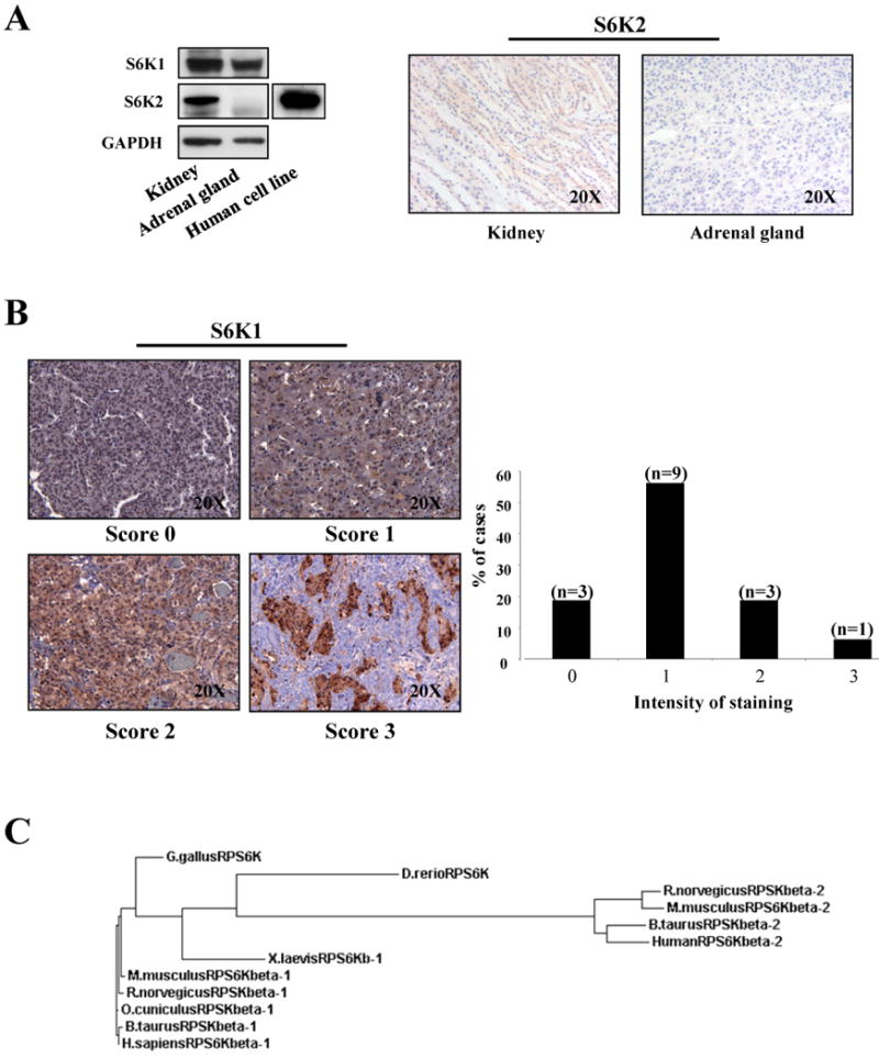
Figure 3. S6K1 and S6K2 proteins expression in human tissues.

A, left panel: western blot analysis on human tissues lysates. As a control we have used a lysate from MCF7 human cell line. Right panel: IHC with anti-S6K2 on normal human tissues. B, staining with anti-S6K on 16 cases of human pheochromocytoma. S6K1 immunostaining was scored as negative (0), weak (1), moderate (2) or strong (3). C, phylogenetic tree indicates the relationship and evolutionary descent of various species based on protein sequence of S6K1 and S6K2. Specifically, RPSKbeta-1 refers to S6K1 protein and RPSKbeta-2 to S6K2.
